
Seeing lungs in 3D: a breakthrough for ICU ventilator care
Erasmus MC is developing 3D lung imaging for ICU patients, helping doctors fine-tune ventilation for every individual.
Published on August 20, 2026
© William Verbakel - Erasmus MC

Team IO+ selects and features the most important news stories on innovation and technology, carefully curated by our editors.
Doctors at Erasmus Medical Center in Rotterdam are developing a new way to monitor the lungs of patients on mechanical ventilation — and this time, they're doing it in 3D. The technology could help medical teams fine-tune ventilator settings for each individual patient, reducing the risk of harm from a treatment that, while lifesaving, can also cause damage.
Patients in intensive care units (ICUs) who can't breathe adequately on their own are given air under pressure through a ventilator. Sometimes the machine takes over breathing completely; sometimes it just lends a hand. As physician-engineer Annemijn Jonkman puts it, diseased lungs behave like an unevenly stretchy balloon — some areas barely expand while others get overstretched. Finding the right amount of support without causing extra harm is delicate work. "Ventilation is damage," she sometimes says, only half-joking.
.png&w=2048&q=75)
Colleague Peter Somhorst explains why the balance is so tricky: too little pressure means insufficient ventilation, too much pressure risks overstretching and damaging lung tissue. To complicate things further, not all parts of the lungs are equally affected by disease. Getting that balance right at the bedside requires a much clearer picture of what's actually happening inside the lungs in real time — which is exactly what Jonkman and Somhorst are working to provide.
Watching the lungs breathe, live
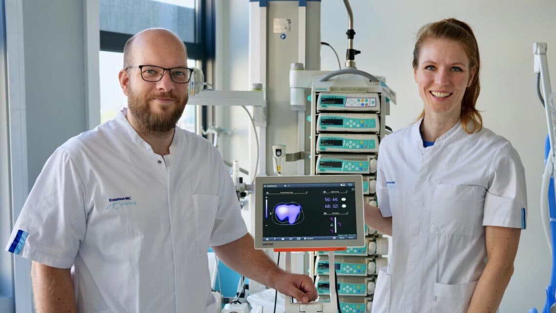
Their tool is called Electrical Impedance Tomography, or EIT. ICU patients wear two sensor bands around their chests, which detect tiny changes in electrical resistance as air moves through the lungs. The system generates a new image up to 50 times per second, allowing clinicians to follow the ventilation process in real time.
What makes the 3D version new is the extra dimension it adds. While the current 2D technology — already in use at Erasmus MC's ICU for 12 years — only shows a horizontal cross-section of the lungs, the 3D version also reveals differences from top to bottom. Jonkman expects this to be especially useful for patients working toward breathing independently again, since it shows exactly how different lung regions are contributing to each breath — valuable information for deciding when it's safe to start reducing ventilator support.
Lessons from the pandemic
The need for this kind of personalized insight became strikingly clear during the COVID-19 pandemic. Somhorst recalls two broad patient types: one group became severely ill quickly, often with a higher Body Mass Index (BMI), and needed higher ventilation pressures; the other stayed ill longer, wasn't overweight, but suffered from blood-clotting problems that hindered oxygen uptake. "There is no single optimal way to ventilate," he says — which is precisely why understanding each patient's lungs individually matters so much.
Preventing ventilator-induced damage, he adds, could mean faster recovery and fewer lingering complaints for patients, fewer complications and shorter hospital stays for healthcare systems, and a quicker return to work and daily life for society at large.
International collaboration
Recognizing that potential impact, the Dutch Research Council (NWO) awarded the project just over €900,000 in funding. Jonkman and Somhorst, together with ICU department head Diederik Gommers, formed a consortium that includes a medical technology company as well as researchers from Carleton University in Canada and the Fraunhofer Institute in Germany. The partners are jointly developing open-source software and algorithms, with the aim of making the technology available for use and further improvement worldwide.